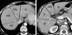
Couinaud Liver Segments in CT Scanner
A. Superior portion of the liver. B. Inferior portion of the liver. CT scans illustrate the Couinaud classification of numbering of liver segments. The longitudinal plane of the right hepatic vein divides VIII from VII in the superior portion of the liver and V from VI in the inferior portion of the liver. The longitudinal plane of the middle hepatic vein through the gallbladder fossa separates IVa from VIII in the superior liver and IVb from V in the inferior liver. The longitudinal plane of the left hepatic vein and fissure of the ligamentum teres separates IVa from II in the superior liver and IVb from III in the inferior liver. The axial plane of the left portal vein separates IVa superiorly from IVb inferiorly and II superiorly from III inferiorly in the left lobe. The axial plane of the right portal vein separates VIII and VII superiorly from V and VI inferiorly in the right lobe. The caudate lobe (segment I) extends between the fissure of the ligamentum venosum anteriorly and the inferior vena cava posteriorly.

Great Site. Very helpful.
cool nice pix 🙂 i like it. May go to vn and see u there Ban.
Good
good!
really helped.. i was confused .. thank you very much…